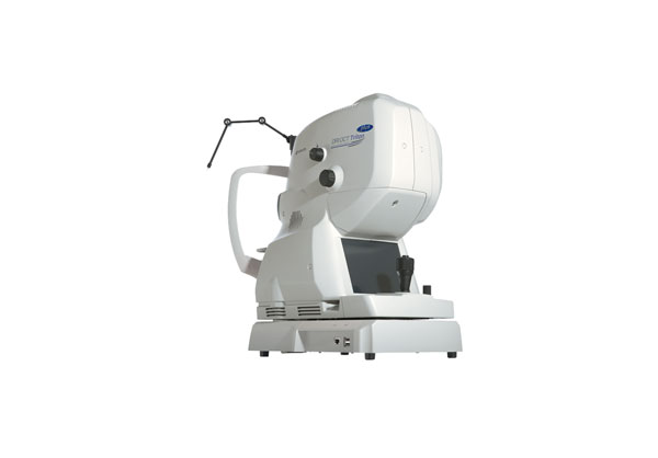
Topcon TRITON /TRITON PLUS
Swept Source adds a new dimension to OCT. The Topcon DRI OCT Swept Source is easy to use, provides
unique clinical information, and has improved my practice. For the first time, we can in-vivo visualize not only
the vitreo-retinal interface but also the cortical vitreous which is important at the time when more and more
therapies are delivered via intra-vitreal injections. Deeper imaging brings choroidal thickness, helping guide
my clinical decisions. Seeing more helps guide my therapy and allows me to treat more effectively. I find
Swept Source OCT an essential tool to look for biomarkers of disease regression or progression.
Prof. P. E. Stanga, Manchester Royal Eye Hospital, Manchester
Description
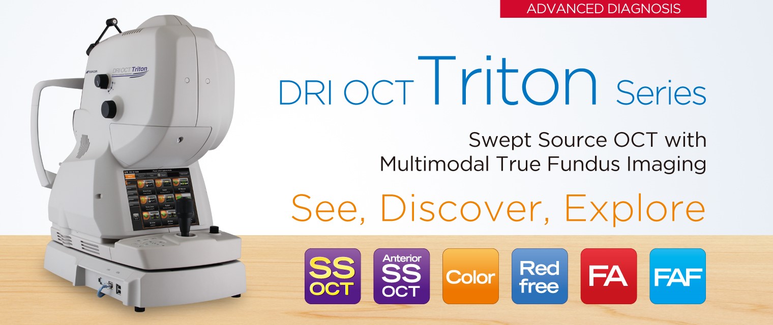
SEE. DISCOVER. EXPLORE.
The diagnostic power of Swept Source OCT
Deep Range Imaging.

Welcome to the new frontier in OCT imaging
The DRI OCT Triton combines the world’s first Swept Source OCT technology with multimodal fundus imaging.
Multimodal All-in-One fundus imaging tool will bring the next level of diagnostic capability to you and your patients.
Unprecedented image quality
Triton’s Swept Source with its extremely fast scanning speed and longer 1,050nm wavelength results in stunningly clear, detailed images, even into the deepest layers of the eye with short acquisition
time. You will not only see the retina and vitreous, but also the choroid and the sclera like never before.
Remarkable diagnostic capability
Seeing deeper makes it possible to have a better understanding of many ocular pathologies, and may
provide the advantage of early disease detection and monitoring. Combined with unique features such as OCT Angiography and En Face imaging, Triton empowers you to take proactive steps to preserve your patients’ eye health.
Greater clinical efficiency
A wealth of automated and intuitive functions, including single-scan captures and the new SMARTTrackTM system, are designed to optimize your practice workflow by simplifying data capture,
analysis, and follow up.
Swept Source OCT deep range imaging
Swept Source technology & 1,050nm wave length
Swept source OCT provides a significant improvement over conventional OCT. Due to the optimized long wavelength scannning light (1,050nm), there is better penetration of the deeper layers of the eye. Furthermore, this scanning light also penetrates better through cataracts, hemorrhages, blood vessels and sclera.
The world’s fastest*1 scanning speed 100,000 A-Scans/second
Approximately twice higher scan speed*2 will brings more scans for a single B scan image, and more informative image supports efficiency and quality of diagnosis.
*1 According to TOPCON survey -Feb. 2015.
*2 Compared with TOPCON SD OCT.

SEE DEEPER. SEE MORE
Proliferative diabetic retinopathy

Source: Prof. P. E. Stanga, Manchester Royal Eye Hospital, Manchester Vision Regeneration (MVR) Lab at N IHR/ Welcome Trust Manchester CRF & University of Manchester
Central serous retinopathy

Source: Prof. P. E. Stanga, Manchester Royal Eye Hospital, Manchester Vision Regeneration (MVR) Lab at N IHR/ Welcome Trust Manchester CRF & University of Manchester

Source: Prof. P. E. Stanga, Manchester Royal Eye Hospital, Manchester Vision Regeneration (MVR) Lab at N IHR/ Welcome Trust Manchester CRF & University of Manchester

Source: Prof. P. E. Stanga, Manchester Royal Eye Hospital, Manchester Vision Regeneration (MVR) Lab at N IHR/ Welcome Trust Manchester CRF & University of Manchester
Pathological myopia

Source: Prof. P. E. Stanga, Manchester Royal Eye Hospital, Manchester Vision Regeneration (MVR) Lab at NIHR/ Welcome Trust Manchester CRF & University of Manchester
Maküler pucker
Image through cataract

A


B

C
Source: Prof. P. E. Stanga, Manchester Royal Eye Hospital, Manchester Vision Regeneration (MVR) Lab at NIHR/ Welcome Trust Manchester CRF & University of Manchester
A B C ) Source: Kazuya Yamagishi, MD (Hirakata Yamagishi Eye Clinic, Japan)

